Section 1 · Project Overview
Treatment and workflow with histological kidney tissue affected by nephrocalcinosis.
Treatment and Work with Histological Kidney Tissue Affected by Nephrocalcinosis
The project documents a complete workflow for handling kidney tissue influenced by nephrocalcinosis — from fixation to microscopic assessment — focusing on preserving tissue integrity and diagnostic value throughout processing.
Fixation
Tissue that is extracted from an organism must be immediately fixed to prevent damage caused by internal and external factors, such as the onset of putrefaction. Shown here is how the tissue was delivered, fully fixed, and prepared before macroscopic examination and assessment.
Macroscopic Assessment and Trimming
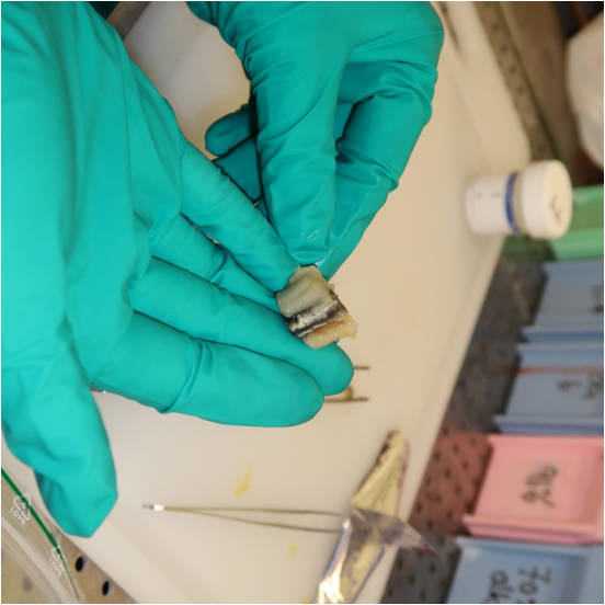
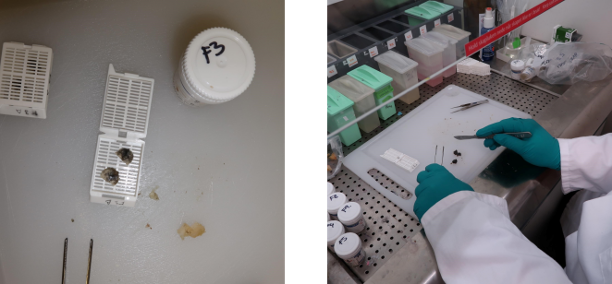
The tissue material is assessed for consistency, color, and size — this evaluation is part of the diagnostic process. The kidney tissue is sliced so that, under microscopy, relevant components can be observed in each section. The slices are oriented and placed in labeled cassettes, with detailed information systematically recorded.
Feeding (Tissue Processing)
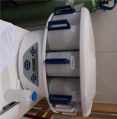
The tissue was processed using the SPIN TISSUE PROCESSOR STP10. This step prepares it for paraffin embedding. For successful embedding, tissue must become permeable to paraffin — achieved through formalin fixation, dehydration, clearing in xylene, and final paraffin impregnation.
Paraffin Embedding
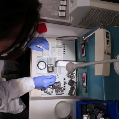
Once permeable, tissue slices can be embedded in paraffin using the MEDITE TBS88 – Paraffin Embedding System. After the paraffin in the chamber has melted, a mold is selected based on tissue size, a small amount of paraffin is poured, and the tissue is properly oriented. The mold is then moved to a cold plate to accelerate solidification while pressing the tissue to ensure the surface adheres. The labeled cassette with ID is attached, and the entire mold is placed on a larger cold plate until fully solidified.
Sectioning
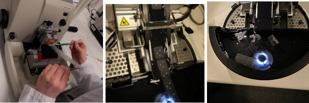
The finished paraffin blocks are trimmed to ensure that each section includes the full tissue surface. Trimming requirements vary per block. Various microtomes — manual or with water slides — are used. Precision and patience are essential for clean, even sections. For disease-affected samples, extra care is taken to avoid tearing and ensure complete component preservation. The sections are floated on a water bath, mounted on slides, and placed on a warming plate for adhesion.
Staining
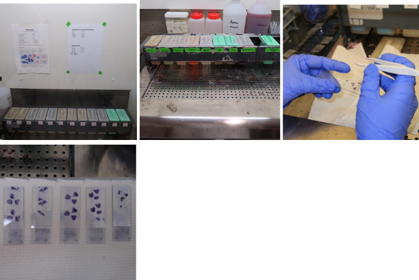
To visualize tissue components under the microscope, an appropriate staining method is chosen. HE/HES is a common stain that differentiates layers and structures, providing clear contrast between various cell and tissue types.
Microscopic Assessment
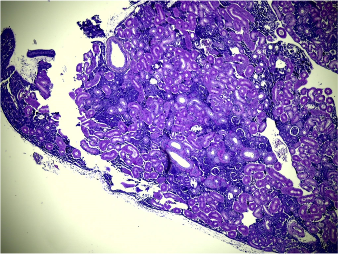
During microscopic examination, normal histology is identified — including tissue architecture, cell types, and characteristic structures. Abnormal findings such as tissue damage, inflammatory or blood cells, and necrosis are also evaluated, as these indicate pathological changes absent in healthy tissue.